One Of The Best Tips About How To Draw Restriction Mapping

We know from the vector map that the vector.
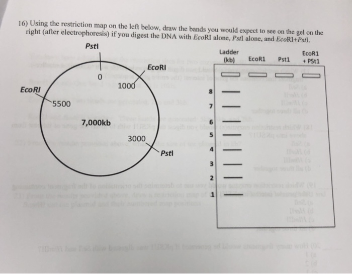
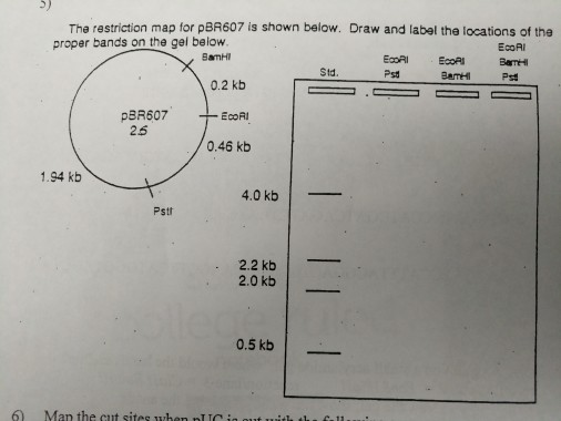
How to draw restriction mapping. Dna cut with ecori + hindiii 350, 300, 200, 50 maps sites for restriction enzymes, a.k.a. Here, two portions of the dna sample are individually digested with. Learn to separate dna on an agarose gel using electrophoresis.
2)you know the total size of the plasmid is 100 kb since the lengths of the fragments in each lane add up to. Compare the λ dna bands on a gel to the. Below is shown an agarose gel of the appropriate digests.
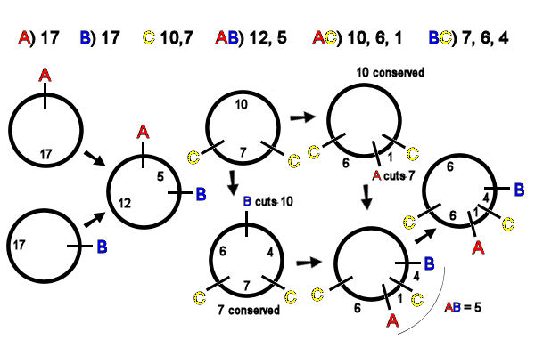
•another way to construct a restriction map •expose dna to the restriction enzyme for a limited amount of time to prevent it from cutting at all restriction sites (partial digestion) •generates. Draw the next site x+y spaces from the 0/start space when. First we know that the total size of the recombinant plasmid must be 1000 bp (the sum of all of the fragments in any lane in the gel).
Place the next cut size x spaces to the right of the 0/start space where x equals the size of the smallest fragments. One approach in constructing a restriction map of a dna molecule is to sequence the whole molecule and to run the sequence through a computer program that will find the recognition. As a first step, you construct a restriction map of the fragment using the enzymes sma i and hin diii.
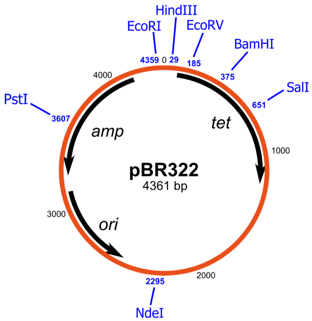
Mapping will be accomplished by performing single and double restriction digests of the plasmid followed by separation of the dna fragments using agarose gel electrophoresis. A restriction map is a diagram that indicates the relative positions of restriction enzyme sites on. Using gel electrophoresis, it's possible to measure size of resulting restriction fragments.
1) cut out a circle of paper and use that to trace another circle on a sheet of paper.


